
How Do X-Ray, MRI, and CT Scans Actually Differ?
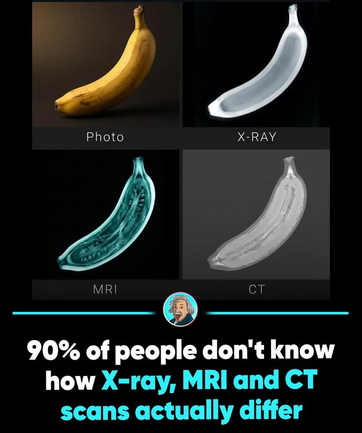
Social media platforms are frequently populated by striking visual graphics, science trivia, and viral clickbait headlines designed to test your knowledge or capture your attention. Among the most popular educational posts circulating online are infographics comparing everyday objects—such as a banana—under different medical imaging technologies like X-rays, MRIs, and CT scans. For anyone curious about medical technology, these viral posts offer a fascinating way to visualize how advanced diagnostic tools “see” inside objects.
Understanding how medical imaging works involves exploring radiology, physics, and clinical diagnostics without relying solely on social media graphics. As radiologists and medical professionals emphasize, each imaging modality utilizes fundamentally different physical principles to capture internal structures. By exploring the facts behind medical scans, practical health awareness, and common misconceptions, you can approach science trivia with clarity and confidence.
The Physics of Medical Imaging Technologies
To understand how X-rays, MRIs, and CT scans differ, it helps to examine the core scientific principles behind each technology.
X-Ray Imaging: Traditional X-rays pass ionizing radiation through an object or body, capturing a two-dimensional shadow image where denser materials block more radiation and appear lighter.
CT (Computed Tomography) Scans: CT scans utilize specialized X-ray equipment combined with computer processing to take multiple cross-sectional images (slices) from various angles, creating a detailed three-dimensional view.
MRI (Magnetic Resonance Imaging): MRIs do not use radiation; instead, they employ powerful magnetic fields and radio waves to excite hydrogen atoms in water molecules, producing exceptionally detailed images of soft tissues.
Practical Tips for Health Literacy and Medical Navigation
Navigating health information online and interacting with modern medical care involves developing a solid understanding of diagnostic tools.
Consult Healthcare Professionals: Always discuss the necessity, purpose, and safety of any recommended medical imaging procedure with your physician or radiologist.
Understand Radiation Safety: When undergoing X-rays or CT scans, doctors use the lowest effective dose necessary to obtain clear diagnostic information.
Prepare for Scans: Follow specific preparation instructions provided by your clinic, such as removing metal objects for MRIs or fasting for certain contrast scans.
Verify Scientific Sources: Cross-reference viral science graphics with accredited medical institutions, radiology associations, and peer-reviewed educational resources.
Interesting Facts About Medical Imaging History
Accidental Discovery: Wilhelm Röntgen discovered X-rays in 1895 while experimenting with cathode ray tubes, forever changing the landscape of medical diagnostics.
Viral Appeal: Educational graphics comparing medical scans of everyday objects spread rapidly across social media because they combine stunning visuals with accessible science trivia.
Nobel Prize Recognition: The development of MRI technology earned key researchers the Nobel Prize in Physiology or Medicine in 2003 for its revolutionary impact on clinical care.
Non-Invasive Diagnostics: Modern imaging allows physicians to examine internal structures, organs, and injuries with zero surgical invasion, vastly improving patient outcomes.
Common Misconceptions About Diagnostic Scans
Navigating online science advice requires distinguishing between educational graphics and clinical realities.
| Myth | Reality | Recommended Alternative |
| “All Medical Scans Use the Same Technology” | X-rays, CT scans, and MRIs rely on completely different physical principles, from radiation to magnetism | Consult a radiologist or physician to understand your specific imaging procedure |
| “Viral Infographics Replace Medical Textbooks” | Social media graphics simplify complex medical physics for quick consumption and visual engagement | Verify scientific facts through accredited medical foundations and radiology resources |
| “MRIs and CT Scans Are Interchangeable” | Doctors choose specific scans based on whether they need to visualize bones, soft tissue, or blood vessels | Follow professional medical orders when diagnostic imaging is required |
| “Radiation from Routine Scans Is Always Dangerous” | Medical imaging radiation doses are strictly controlled and outweighed by diagnostic benefits | Discuss any safety concerns directly with your healthcare provider |
Expert Recommendations for Patient Education
Radiologists, physicians, and healthcare educators emphasize that understanding medical technology empowers patients during clinical visits. Relying solely on social media memes for medical education can lead to misunderstandings about how diagnostic tools operate.
Keeping your health records, imaging history, and physician notes organized ensures you maintain clear communication with your medical team. Prioritizing verified professional guidance provides the safest foundation for health literacy.
Conclusion
The idea of viewing a banana through X-ray, MRI, and CT scan lenses makes for a brilliant and engaging viral post, demonstrating the diverse capabilities of modern medical imaging. By relying on verified science, supporting health literacy, and filtering out sensationalized clickbait, you can appreciate the incredible physics that help doctors diagnose and heal every day.
Frequently Asked Questions
1. What is the main difference between an X-ray and a CT scan?
An X-ray provides a single two-dimensional shadow image, whereas a CT scan takes multiple X-rays from various angles to create detailed 3D cross-sectional slices.
2. Do MRIs use radiation?
No, MRIs do not use ionizing radiation; instead, they utilize powerful magnets and radio waves to generate detailed images of soft tissues.
3. Why do viral posts use everyday objects like bananas for medical comparisons?
Visualizing advanced technology on familiar objects makes complex physics accessible, captivating user attention and driving social media shares.
4. When would a doctor choose an MRI over a CT scan?
Doctors typically choose MRIs for superior visualization of soft tissues, brains, ligaments, and spinal cords, while CT scans are often favored for bones and emergency trauma.
5. Are medical imaging scans safe?
Yes, medical scans are performed under strict safety guidelines where the diagnostic benefits vastly outweigh the minimal risks associated with controlled radiation or magnetic fields.
6. Can metal objects be worn inside an MRI machine?
No, the powerful magnetic field of an MRI machine will attract metal objects, making strict screening mandatory before entering the scanning room.
7. How long did it take to develop MRI technology?
The foundational physics for MRI were developed over several decades through the mid-to-late 20th century before becoming clinically viable in the 1980s.
8. Why is medical accuracy important in science communication?
Accurate science communication prevents public confusion, reduces medical anxiety, and ensures people understand the true purpose of diagnostic tools.
9. When should I ask my doctor about imaging safety?
You can ask your physician about imaging safety at any time before a procedure, especially if you are pregnant, have metal implants, or feel anxious.
10. How can I spot reliable science information online?
Look for content backed by peer-reviewed research, accredited scientific institutions, medical boards, and qualified healthcare professionals.